SIXMA GROUP
Research

Sixma Group

Research

Research

Research programmes

We present four interlocking programmes that connect enzymatic mechanism with chromatin and repair biology.
01

Ubiquitin conjugation and deubiquitinating enzymes: decoding activity and specificity

How do DUBs and ubiquitin ligases recognise specific substrates and chain architectures, and how does that determine DNA repair outcomes?

Ubiquitin modifications come in many chain types and architectures; the same modification can signal different outcomes depending on context. We determine structures of DUBs bound to defined ubiquitin conjugates and measure cleavage kinetics to understand how enzymes discriminate between polyubiquitin and monoubiquitin adducts. This mechanistic view reveals how DUBs can be regulated allosterically and by binding partners.

Beyond in vitro enzymology, we integrate quantitative assays in cells to map how DUB activity changes at sites of replication stress and DNA damage. These data allow us to connect biochemical mechanism to functional consequences for genome stability and to explore how DUBs become candidate targets for therapeutic modulation.
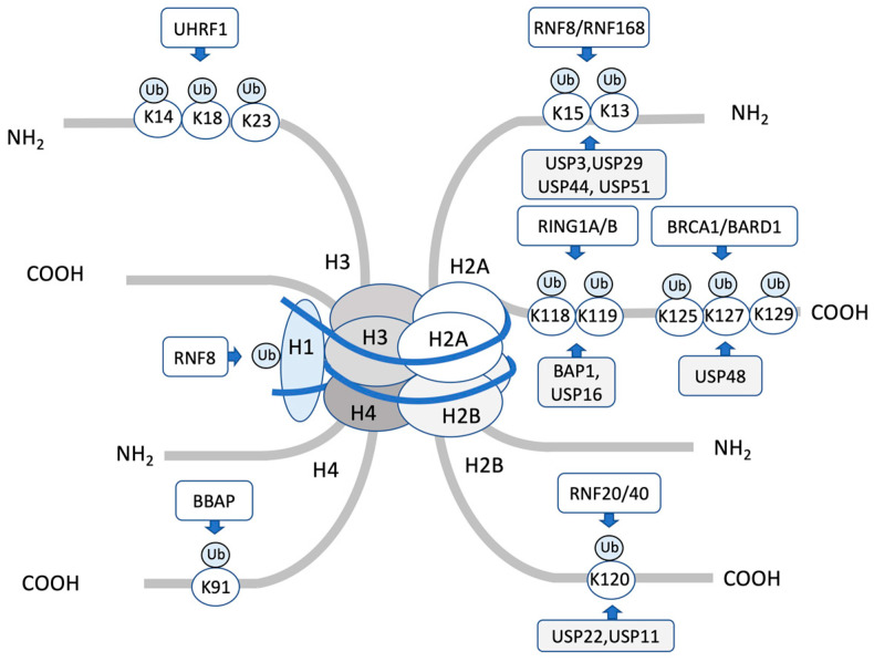
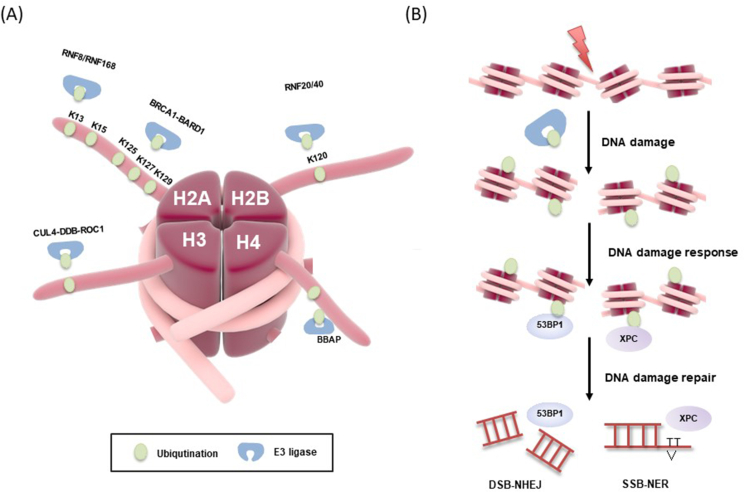
Figure 1 from 'DNA Damage Response Regulation by Histone Ubiquitination' (Int. J. Mol. Sci., 2022) — schematic overview of histone ubiquitination events at DNA double-strand breaks and roles of E3 ligases and DUBs in repair-pathway choice (used to illustrate substrate/chain architecture recognition and functional consequences).
02

Chromatin regulation of the DNA damage response

How does histone ubiquitination and deubiquitination change chromatin to permit repair factor access?

Damage signalling remodels chromatin by adding and removing ubiquitin marks on histones such as H2A and H2AX. We study the structural basis for recognition of histone ubiquitination states by effectors and how specific DUBs remove those marks. Our structural snapshots and biochemical reconstitutions explain how chromatin context modulates enzyme function.

By combining nucleosome reconstitution with enzymology and cell biology, we probe how ubiquitin‑dependent chromatin states influence repair pathway choice and checkpoint signalling. This work draws on collaborations with chromatin and repair specialists to place molecular mechanisms in a physiological context.

Figure 3 from 'Histone post-translational modification and the DNA damage response' (review, 2022) — illustration of histone ubiquitination marks, readers and erasers at DSBs and how chromatin is remodelled to permit repair factor access.
03

PCNA ubiquitination and replication‑associated repair

How is PCNA ubiquitylation regulated and reversed to control lesion bypass and restart?

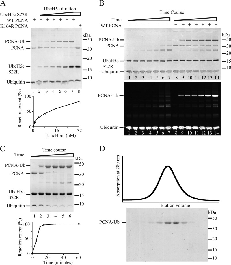
PCNA ubiquitylation is a central switch during replication stress, controlling translesion synthesis and template switching. We focus on the enzymatic mechanisms that alter PCNA ubiquitination status, including the action of USP1/UAF1 and other DUBs, combining structural insight with kinetic characterisation.

Understanding how cells temporarily enrich or remove specific PCNA ubiquitylation states informs on how replication forks cope with lesions and how dysregulation can lead to genomic instability in cancer. We pursue biochemical reconstitution of polyubiquitinated PCNA substrates and interrogate how enzymatic preferences translate to cellular repair outcomes.

Figure 1 from 'Intrinsic Flexibility of Ubiquitin on Proliferating Cell Nuclear Antigen (PCNA) in Translesion Synthesis' (PMC3493961) showing structural/biochemical data on ubiquitinated PCNA relevant to regulation of PCNA ubiquitylation and downstream lesion bypass.
04

BRCA1 interactions and the architecture of repair assemblies

Which BRCA1 interfaces are essential for partner recruitment and homologous recombination competence?

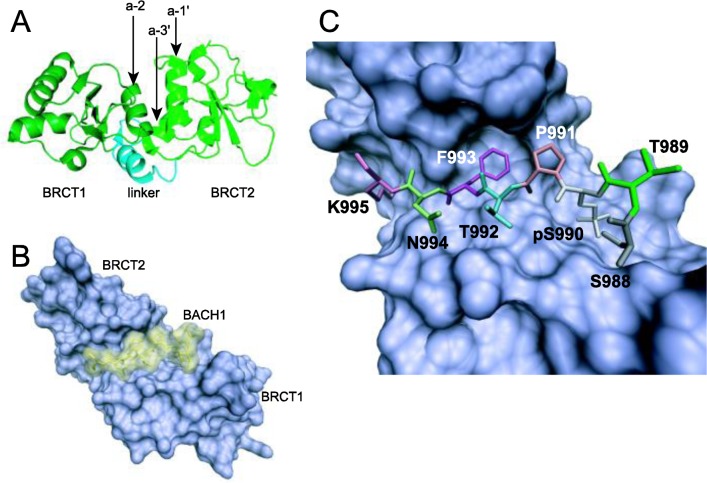
We map protein–protein interfaces involving BRCA1 BRCT domains using high‑resolution biochemical and structural approaches. Precise interaction maps help interpret how patient variants affect BRCA1 function and homologous recombination efficiency.

This programme links structural mapping with cellular assays of homologous recombination and with functional genomics, yielding an integrated picture useful for variant interpretation and for identifying nodes that could be targeted to modulate repair in cancer.

Figure 4 from 'Structure-Function of the Tumor Suppressor BRCA1' (review, PMC3380633) — structural view of BRCT tandem repeats and peptide recognition pockets, useful for understanding BRCA1 partner interfaces and binding architecture relevant to repair-assembly function.